-
Farewell KCG
06/01/2026
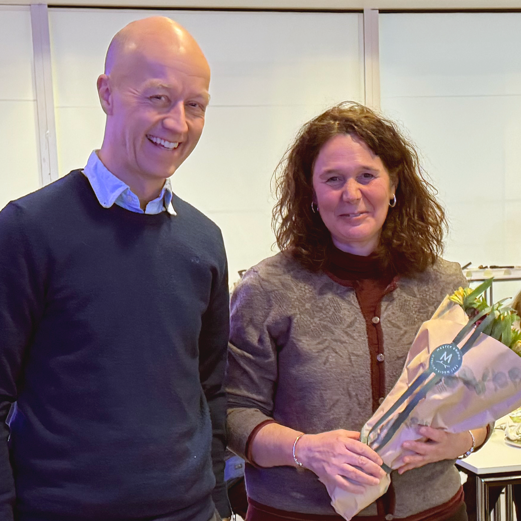 Tarjei Hveem and Francesca Micci
🎉 Celebrating 16 Years of Cancer Cytogenetics (and a lot of laughter) at ICGI 🎉
In January 2026, we bid a heartfelt farewell to the Cancer Cytogenetics (KCG) section
and thank them for 16 years as an integral part of ICGI. With deep expertise and a
highly specialised laboratory, the section translates complex genomic findings into
clinically meaningful insights, many of which have a direct impact on personalised
treatment. KCG have been a key contributor to the institute’s extensive portfolio of
peer-reviewed scientific publications.
📍 A practical update: KCG is transferring within Oslo University Hospital, but not
moving location — you’ll still find them on the 5th floor of the Oslo Cancer Cluster
building. Their test requisition forms for analyses will now be available on
ous.labfag.no
And yes — the rest of ICGI will miss hearing “karyograms” and “fusion genes” in
everyday conversation. But beyond the terminology, we will miss the people most:
the highly skilled, creative, and good-humoured colleagues who have enriched ICGI
with deep expertise and many memorable moments.
💙 Thank you for 16 fun, meaningful, and impactful years.
As KCG moves to its new organisational home at the Department of Pathology, Clinic
for Laboratory Medicine, Oslo University Hospital, their essential work in cancer
diagnostics and personalised treatment remains firmly established. Our organisational
“marriage” may have come to an end, but our friendship — and collaboration — certainly
continues.
-
Response to letter Re: Surgical stage in the era of molecular profiling of endometrial cancer
01/01/2026
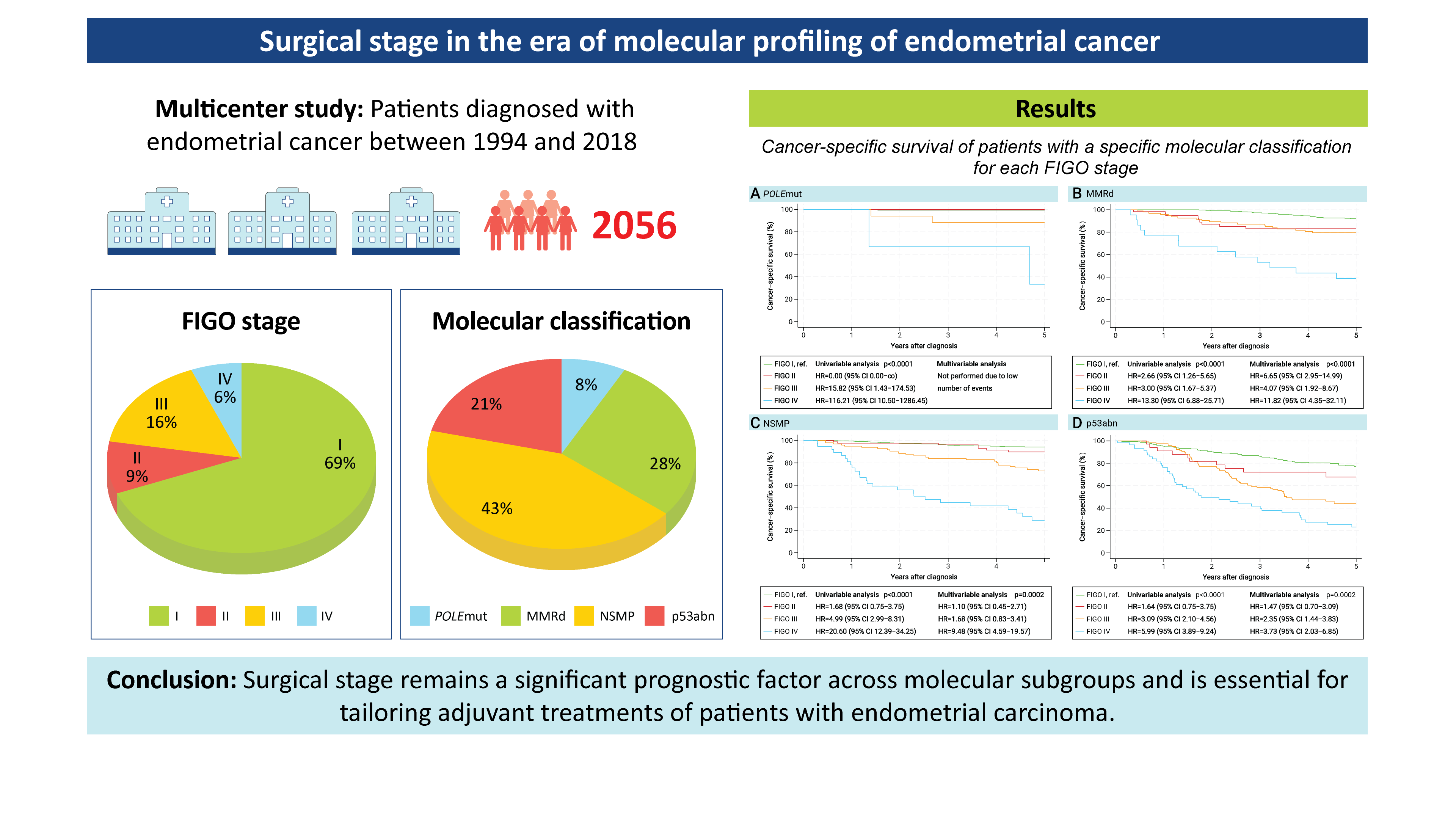 Graphical abstract for the article
We have just published a new study in the European Journal of Cancer✨📘, and are delighted to highlight the work behind this important contribution to the field.
This multicenter study includes more than 2,000 endometrial cancer patients from 11 European centers, making it one of the largest collaborative efforts of its kind. 🤝🌍
In this project, we explored how FIGO staging relates to molecular classification in endometrial cancer. 🔍We found that molecular classification complements—but does not replace—traditional surgical staging.
Our findings help establish the role of FIGO staging in the era of molecular profiling, leading to safe integration of these decisive disease characteristics for more precise risk assessment and improved patient care.
A big congratulations to all co-authors and partners for their impressive work and dedication.
Find the full-text article through this link.
-
FRIPRO funding for groundbreaking research on digital pathology and artificial intelligence (AI)
15/06/2025
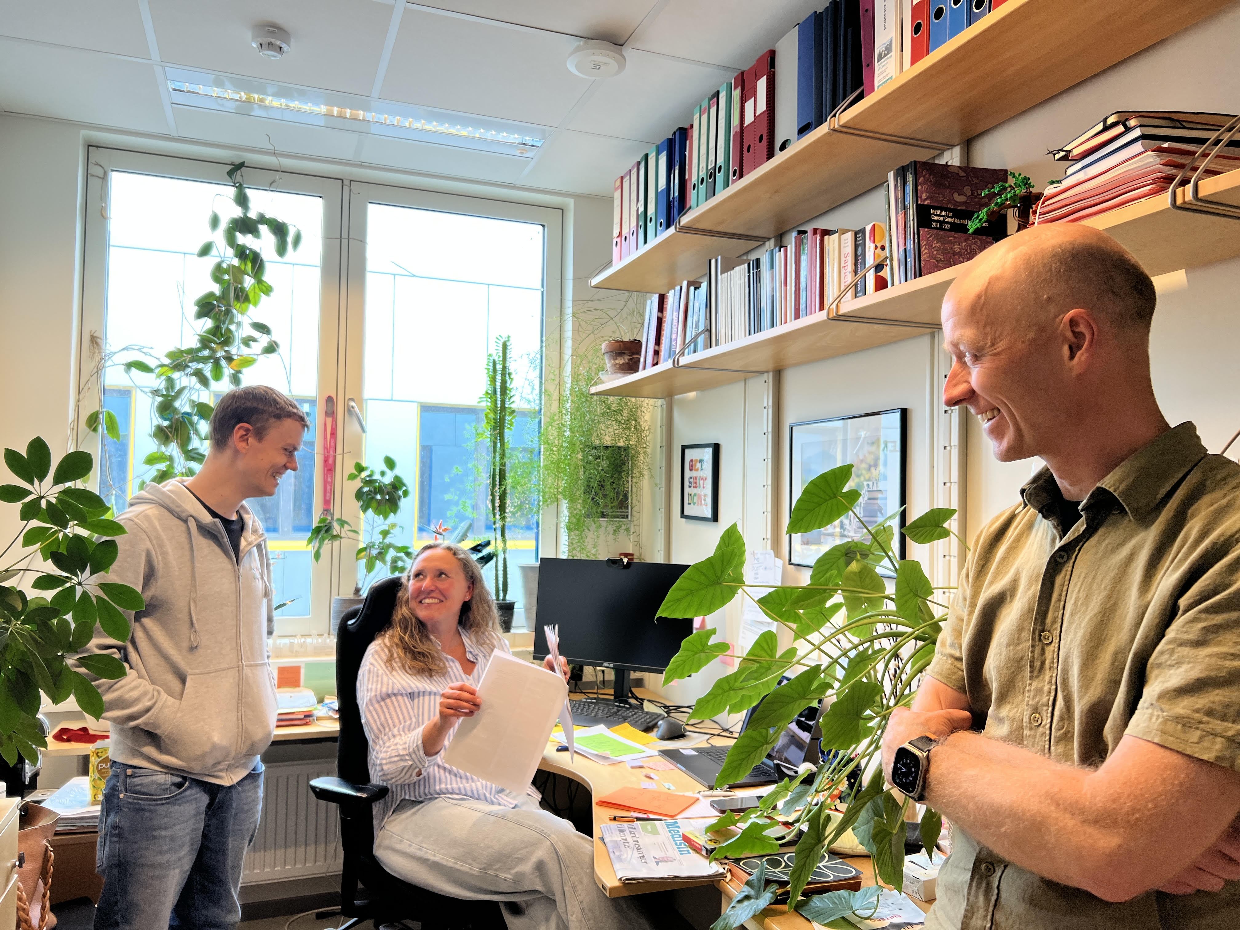 Andreas Kleppe, left, conveying the good news to his colleagues Hanne Askautrud and Tarjei Hveem.
Research Director Andreas Kleppe at the Institute for Cancer Genetics and Informatics (ICGI) has been awarded funding from the Norwegian Research Council's prestigious FRIPRO scheme for the project ENDPATH – End-to-End Pathology. The project aims to develop a new type of imaging system for digital pathology and use the more detailed images to train precise and reliable artificial intelligence (AI) to predict the disease progression of cancer patients, initially patients with prostate cancer.
Unlike current imaging systems that are adapted for visual assessment by humans, the new system will be adapted for automatic analysis with artificial intelligence.
ENDPATH – a boost for digital pathology
Digital pathology is currently used in hospitals across Norway and becoming common in many European countries and elsewhere, opening up new opportunities in diagnostics, research and artificial intelligence. Pathologists make diagnoses and contribute to the choice of treatment, often based on H&E-stained tissue samples. For almost 150 years, pathologists have analyzed tissue samples using this staining method and a microscope. The imaging systems available in digital pathology are designed to provide pathologists with similar images. Most AI models developed for digital pathology are trained using the same images, but both the staining and the imaging system limit the information contained in each image.
The ENDPATH project removes these limitations by developing an imaging system that scans images of tissue samples without staining and uses all the properties of light - amplitude, phase and polarization. This provides richer data for the development of AI models, and facilitates more precise and robust tools in future digital pathology. The project will exemplify this by developing AI models that will indicate the risk of disease progression in prostate cancer from images of biopsy samples taken during active surveillance of assumed low-risk patients. Although the new type of images may be difficult for pathologists to interpret directly, they can be converted to look like the H&E-stained tissue samples that pathologists are experts at evaluating, thus the new imaging system could potentially replace current systems in the future without impacting the pathological assessments performed in clinical practice.
Fierce competition – high quality
FRIPRO is a national funding scheme for groundbreaking research known to be highly competitive. Only applications that receive top marks on all assessment criteria are eligible for funding. The fact that Andreas Kleppe and this project are among the selected few, is clear proof of both quality and originality. “At FRIPRO, we are willing to invest in the bold research that has the potential to make significant advances in the field, even if it also has a significant risk of failure,” the Norwegian Research Council argues. The ICGI at Oslo University’s Cancer Clinic has ample experience within digital imaging and AI, and the research group is eager to explore new horizons and shape the next generation of digital pathology.
Read more about the project on Oslo university Hospital's Research website
-
EUnetCCC workshop at OUS
27/02/2025
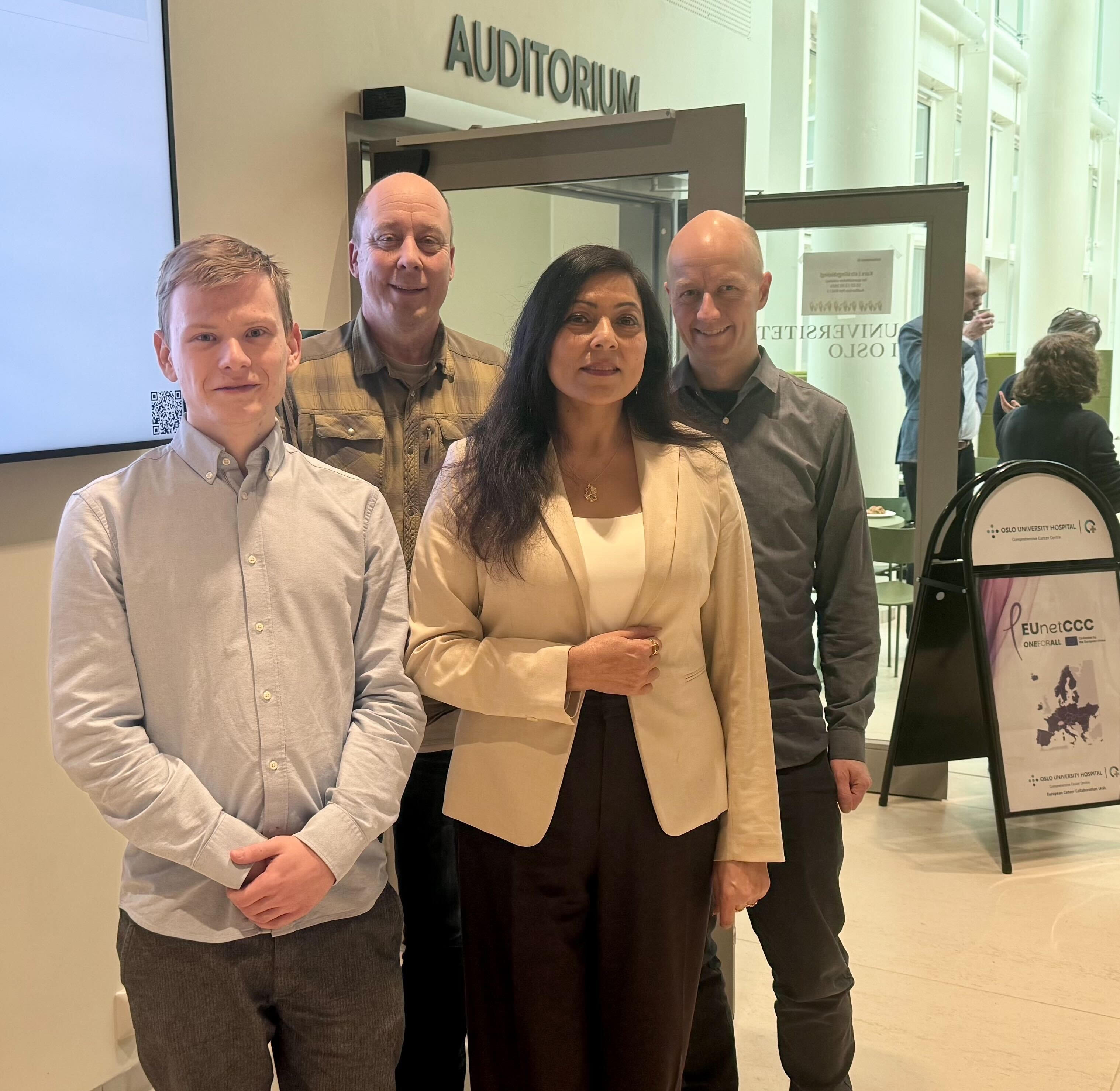 ICGI employees involved in the EUnetCCC project. From left: Marcin Soja, John Arne Nesheim, Rajni Kumar and Tarjei S. Hveem
EUnetCCC WP8 Task Lead Team Workshop is taking place on 27th-28th February 2025 at Oslo University Hospital, Norway.
ICGI is contributing significantly to the EUnetCCC project
This is a major EU initiative to enhance collaboration among European Comprehensive Cancer Centres (CCCs). Oslo University Hospital (OUS) leads Work Package 8, involving 31 countries, 96 institutions, and 60 patient care facilities.
Rajni Kumar is leading this effort at ICGI as the technical project manager and developer. This week, web developer Marcin Soja joined her in their quest to develop web solutions to support collaborative frameworks and toolkits for the network. Welcome, Marcin!
The workshop themes are a range of initiatives, including:
- Enhancing personalized cancer prevention strategies
- Advancing precision cancer diagnostics
- Improving the use of clinical data for research & monitoring
- Developing comprehensive survivorship programs for cancer patients
- Strengthening governance, organization, and leadership in cancer care
The project is aiming to ensure that by 2030, 90% of eligible patients in Europe will have access to high-quality care at Comprehensive Cancer Centres.
-
How to become a world leader in AI and cancer treatment
11/02/2025
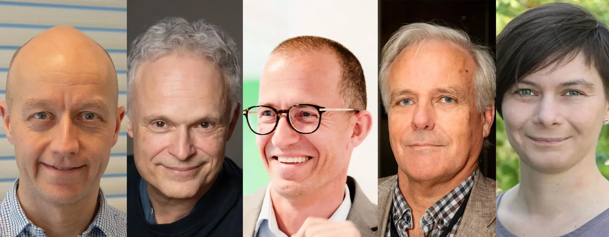 The co-authors of the article in Dagens Medisin and Oslo Cancer Cluster, from left; Tarjei Sveinsgjerd Hveem, Ole Christian Lingjærde, Ketil Widerberg, Sigbjørn Smeland, and Manuela Zucknick.
The Institute for Cancer Genetics and Informatics is pleased to contribute to the ongoing discussion about leveraging AI for improved cancer treatment. Our Interim Director, Tarjei Sveinsgjerd Hveem, recently co-authored an article highlighting Norway's unique health data resources.
Combining our extensive health registries with advanced AI models is the goal of a proposed project called NEXTMAP, which aims to enhance cancer prevention, diagnostics, and treatment. This collaborative effort brings together experienced researchers, companies, and hospitals, pooling a wealth of expertise in cancer research and informatics.
We're grateful to be part of this initiative and ICGI remains committed to supporting this important work by building on the valuable experience of all partners involved.
Read the article in norwegian on dagensmedisin.no, or
in english on Oslo Cancer Cluster's website
-
AI to improve cancer care
15/01/2025
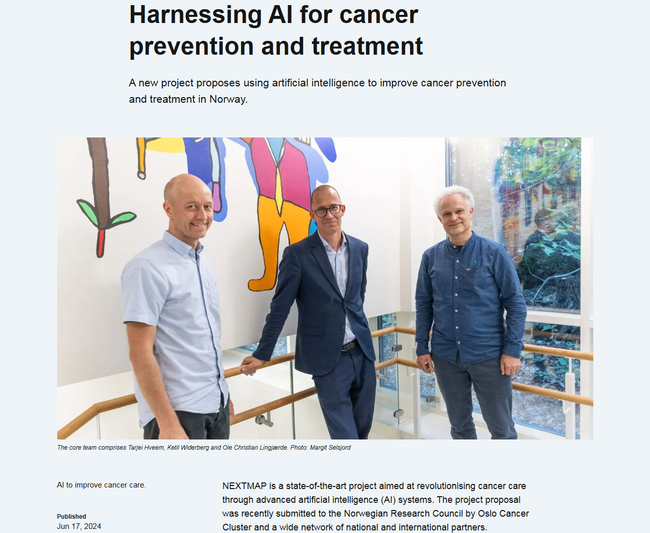 Screenshot from Oslo cancer cluster's website
Project proposal submitted
The first weeks of 2025 has involved late night discussions, long-distance video meetings, and detailed manuscript editing for the application team of NEXTMAP.
On January 15th 2025, the detailed project proposal was submitted to the Norwegian Research Council.
Alongside ICGI, one of the partners in the project is Oslo Cancer Cluster, and their article from the first proposal submittance describes the initial purpose of the project.
-
Prognostic and therapeutic implication of molecular classification including L1CAM expression in high-risk endometrial cancer.
16/11/2024
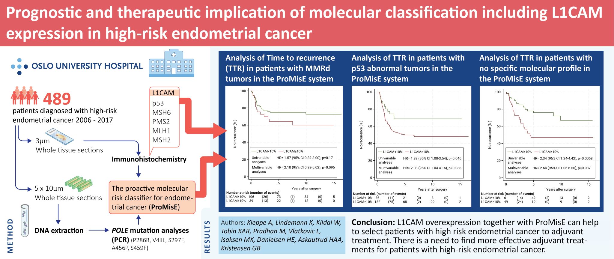 Graphical abstract for the article Prognostic and therapeutic implication of molecular classification including L1CAM expression in high-risk endometrial cancer.
Our article published in the January issue of the journal Gynecologic Oncology, sheds new light on the role of L1CAM, in high-risk endometrial cancer. The article is the result of a collaboration between the Institute of Cancer Genetics and Informatics and the Department of Surgical Oncology, Section for Gynecological Oncology, at Oslo University Hospital, supported by a grant from the Norwegian Cancer Society.
Highlights
- Clearer role of molecular classification and L1CAM in high-risk endometrial cancer.
- ProMisE independently predicted time to recurrence, not cancer-specific survival.
- Patients with POLE mutated tumors had an excellent prognosis.
- L1CAM overexpression was a strong, independent marker for recurrence and survival.
- L1CAM overexpression was related to distant recurrences for the p53 and NSMP group.
Since L1CAM is an additional adverse factor in the p53 abnormal and NSMP groups. These groups need special attention in studies intensifying adjuvant treatment.
The team aims to improve the prognoses and treatment methods for patients with endometrial cancer. The article was opublished online in November 2025.
Find the full-text article through this link.
-
The European Congress of Pathology 2024
25/09/2024
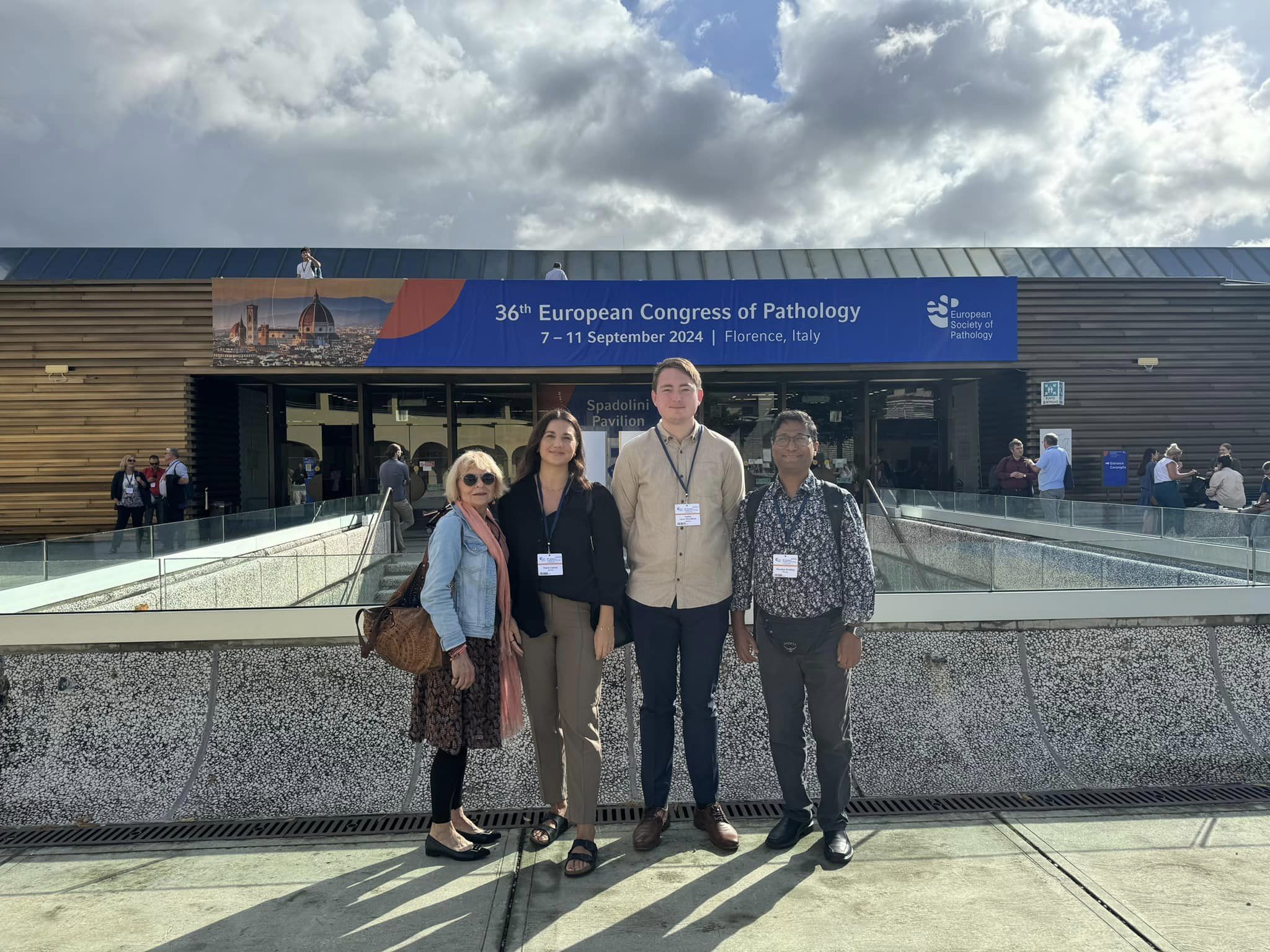 The delegation from ICGI at the European Congress of Pathology in Florenze, Italy From left: Ljiljana Vlatcovic, Maria Isaksen, Audun Ljone Henriksen, and Manohar Pradhan.
"TLS-positive patients had a lower risk of recurrence, especially in tumors with MMR deficiency", said our skilled pathologist, Dr Manohar Pradhan, when he presented research on the prognostic value of tertiary lymphoid structures (TLS) in endometrial carcinoma. The study involves 1,228 patients at Oslo University Hospital, and is a collaboration with the Department of Gynecological Oncology, OUS.
Dr. Pradhans's session was one of 184 featured at the 36th European Congress of Pathology (ECP), which attracted over 5,700 participants from 100 countries. Among them, a delegation from our institute appreciated the opportunity to contribute to and learn from the ongoing advancements within the field of pathology. Additionally Audun Ljone's poster "Improving histopathological screening of colorectal polyps using deep-learning" was on display at the e-poster terminals at the confence venue. ECP was arranged in Florence 7 - 11 September 2024.
We look forward to seeing how insights presented at this year's ECP will contribute to future research and clinical practice.
.
-
Inclusion of several analyzes is beneficial for prostate cancer patients in active surveillance
15/07/2024
Dr. Karolina Cyll at the Department of Cancer Genetics and Informatics (IKI) at Oslo University Hospital and Professor Erik Haug at Vestfold Hospital thank 558 people from Vestfold for their contribution to cancer research. Permission to use data is at the heart of their research paper published in the renowned medical journal British Journal of Cancer (BJC) in July 2024.
.
 Photo from @pexels.com.
Prostate cancer is one of the most common forms of cancer among men worldwide, and around 5,000 Norwegians are diagnosed with this disease each year.
Identify, at an earlier stage, patients who have en increased risk of developing agressive disease
Haug and Cyll show that in addition to PSA and other conventional analyses, it is possible to identify those patients who have an increased risk of developing aggressive disease earlier if DNA ploidy analysis and PTEN status are included in the monitoring protocol. By following this advice, active treatment can be initiated earlier for almost half of the patients who eventually end up needing treatment according to current recommendations.
Read the article (in Norwegian), through this link.
.
-
Dr. Ole-Johan Skrede Successfully Defends his Doctoral Thesis
26/04/2024
We extend our congratulations to Dr. Ole-Johan Skrede for successfully defending his doctoral thesis titled "Selected Studies on the Application of Histological Image Analysis in Cancer Diagnostics Using Deep Learning" on Friday, April 26, 2024. The dissertation took place at the Department of Informatics, Faculty of Mathematics and Natural Sciences, in the namesake's Ole-Johan Dahle’s House.
.
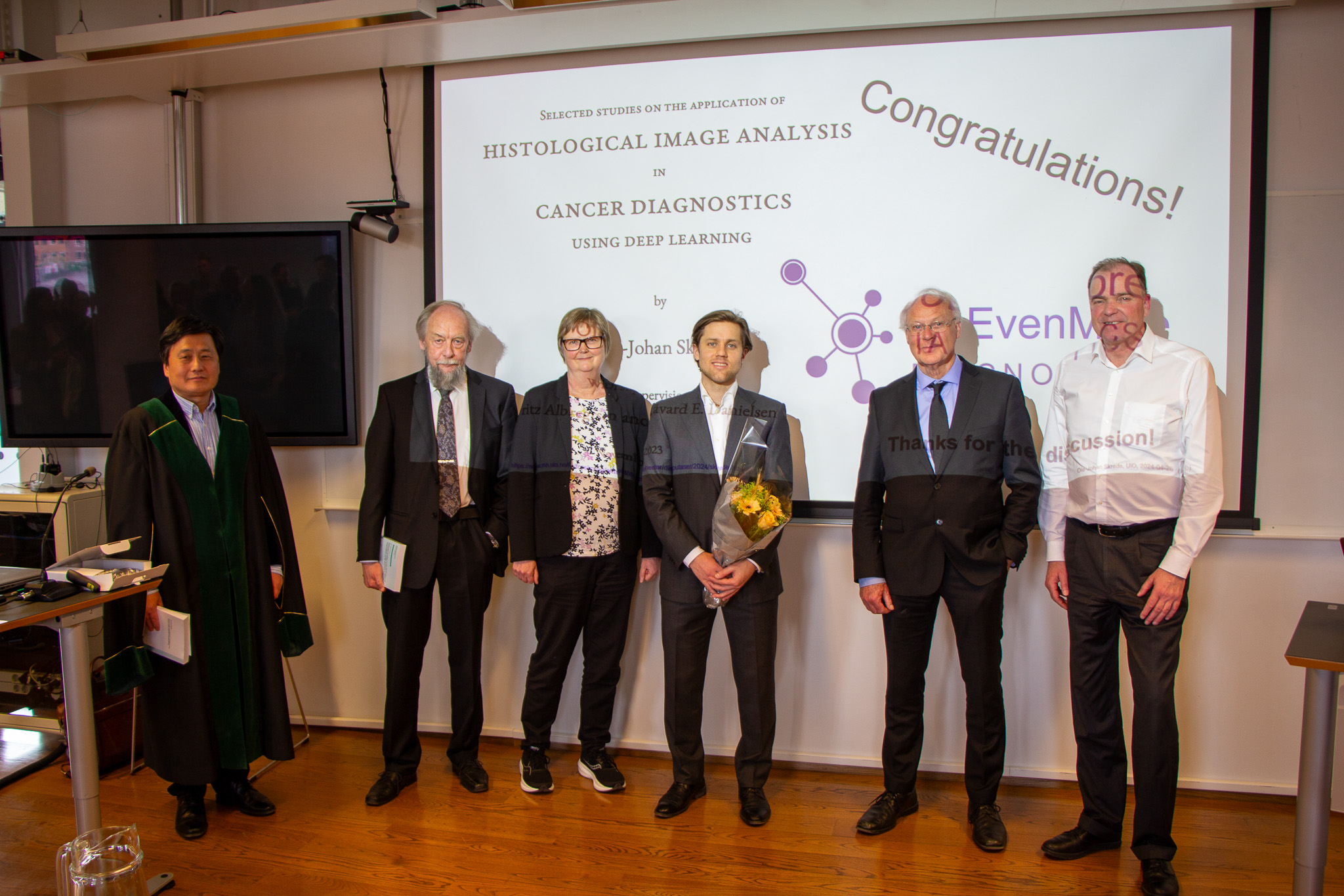 From left: Xing Cai, Supervisor Fritz Albregtsen, Anne Solberg, Ole-Johan Skrede, Anders Lundevold and Paul J van Diest Foto: Petter Bjørklund/UiT
.
From left: Xing Cai, Supervisor Fritz Albregtsen, Anne Solberg, Ole-Johan Skrede, Anders Lundevold and Paul J van Diest Foto: Petter Bjørklund/UiT
.
Dr. Skrede's research focuses on advancing cancer diagnostics through the application of deep learning techniques to analyze histological images. One significant outcome of his work is the development of a method to estimate the prognosis of patients who have undergone colorectal cancer surgery. This innovative approach involves digital microscope image analysis to identify cancerous regions and assess their severity. By training deep learning models on tissue sections from approximately 2,500 patients, Dr. Skrede and his colleagues have developed a deep learning model that enhances the accuracy of prognosis predictions, leading to better stratification of patients to adjuvant chemotherapy after surgery. The research team has rigorously evaluated this methodology on over 1,000 patients to demonstrate its validity and usefulness in clinical practice. Notably, the new method allows identification of substantially more patients that could be spared from unnecessary adjuvant therapy.
.
Dr. Skrede's doctoral thesis comprises three papers published in high-impact journals, significantly contributing to medical research and the recent DoMore project, an ICT Lighthous Project supported by the Research Council of Norway. The papers highlight the integration of deep learning with traditional pathological markers to optimize treatment for patients suffering from colorectal cancer, the possibility to automatically segmented any type of tumor, perhaps even rare types not included in the model development, as well as laying the foundation for better design of deep learning studies in cancer diagnostics and beyond.
.
Before defending his thesis, Dr. Skrede presented a trial lecture at the same venue, on the subject: “Foundation models in cancer research”.
.
The adjudication committee
- Professor Paul J van Diest, Department of Pathology, University Medical Center Utrecht, the Netherlands
- Professor emeritus Arvid Lundervold, Department of Biomedicine, University of Bergen, Norway
- Professor Anne Solberg, Department of Informatics, University of Oslo, Norway
Supervisors
.
Ole-Johan Skrede's supervisors throughout his doctoral journey have been Professor Emeritus Fritz Albregtsen at the Department of Informatics, UiO, Norway, and the late Professor Håvard E. Danielsen, at the Institute for Cancer Genetics and Informatics (ICGI), Oslo University Hospital, Norway.
.
We are so grateful Ole-Johan will continue his research at ICGI, being an important contributor to many of our most prestigious projects.
We extend our gratitude to the committee members for their invaluable insights and to Professor Xing Cai for chairing the defense
.